|
FOUR BASIC STEPS OF MUSCLE CONTRACTION CYCLES |
|
1.attach. of cross-bridge to thin filament 2.movement of X-bridge => prod. tension in the thin filament 3.detach. of the X-bridge fr. thin filament 4.Energizinng the cross bridge so it can again attach to a thin filament & repeat the cycle |
|
POWER STROKE |
|
-ATP hydrolysis puts myosin in a high energy "cocked" state (like a streatch skin)
-Binding of myosin -> actin releases energy -release = conformational change in myosin = "the power stroke"
-result = actin is pulled toward center of sarcomere |
| CYCLE THAT MOVES MYOSIN & HELPS BIND TO ACTIN |
|
-Binding of myosin --> actin needs ATP --> ADP to "energize" -Movement of myosin attach. to actin -detach. of myosin fr. actin (needs ATP Binding) -repeat |
| ACTION POTENTIAL IN SMOOTH MUS. |
|
| ACTION POTENTIAL TRIGGERS Ca FLOW INTO CYTOPLASM |
|
- AP triggers the DHP receptor (voltage-sensitive) - DHP bound to raynodine receptors -ryanodine receptor channel opens Ca2+ |
| AUTONOMIC NERVER INNERVATION OF SMOOTH MUS. |
|
-no motor end plate - Can depolarization/hyperpolarization - swollen regions in autonomic neurons varucosites realse neurontransmitter - Smooth Mus. can slao respond to hormones |
| CARDIAC MUSCLE |
|
| CHAR. OF MUS FIBERS |
+= amt. of SR present in MUS type |
| CHAR. OF MYOSIN |
|
-made of bundles of myosin motors -is actually apolymer of myosin molecules, each of which has a flexible cross-bridge that binds ATP and actin - |
|
CROSS BRIDGE CYCLE (X-bridge) |
|
1.The myosin-bind. site on actin = avail. so the energize cross-bridge binds 2.The full hydrolysis and departure of ADP + Pi causes the flexing of the bound cross bridge 3. Binding of a "new" ATP to the X-bridge uncouples the bridge 4. Hydrolysis= the bound ATP energizes or "re-cocks" the bridge |
| CROSS-BRIDGE CYCLING IS THE SAME AS SKELE MUS |
|
|
Ca2+ IONS PT. 2 |
|
- Regulates myo-actin cycle (X-bridge) - Levels of Ca2+ det. if myo can bind to actin - 2 proteins= troponin + tropomyosin = help regulate process - LOW Ca2+: tropomyo. covers binding sites on actin = prevents myo binding = troponin hods tropomyo in place - HIGH Ca2+: Ca2+ binding on troponin = relaxes binding to tropomyo = tropomyo moves & uncovers binding sites on actin |
| DIFFERENCE BETWEEN NEUROMUSCULAR JUNCTIONS AND REGULAR NEURAL SYNAPSES |
One EPP results in action potential
|
| DIFFERENT BODY REGIONS HAVE DIFFERNT SIZES OF MOTR UNITS |
|
| DIFFERENT SKELETAL MUSCLE CHARATERISTICS |
|
| EXCITATION- CONTRACTION IN CARDIAC MUS. |
|
| FUNCT. OF ATP IN SKELE MUS. CONTRACT. |
|
1. Hydrolysis of ATP by myosin energizes the X-bridges, providing energy for force gen. 2. binding ATP to myo = dissassociates x-bridges bound to actin = allows bridges to repeat cycle of activity 3. hydrolysis of ATP by the Ca2+ -ATPase in the Sacroplas. reticul. provides energy for the active trans. of Ca ions into the reticu. = lowers cystolic Ca to prerelease levels = ends the contraction = the mus. fiber can relax |
| HOW DO ELECTROCHEM. SIGNALS TRANSLATE TO MUS. MOVEMENTS? |
|
- action potential (AC) is initiated in mus. mem. - delay b/t AP and contract - delay due to time need to release Ca2+
|
|
HOW NEUROMUSCULAR JUNCTION WORKS PT 1 |
|
1. synaptic cesicle fusion (triggered by Ca2+) in motorneurons releases acetlcholine 2. on the membrane of the mus. fiber, acetylcholine receptors respond to acetylcholine binding by inc. Na+ entry into the fiber, causing a graded depolarization. 3. the graded depolariz. typically exceeds threshold for the nearby voltage-gate Na+ and K+ channels = so an action potential occurs on the mus. fiber |
| INPUTS INFLUENCING SMOOTH MUS. CONTRAT. ACTIVITES |
|
1. spontaneous elect. activity in the plasma mem. of the mus. fiber 2. neurotansmitters released by autonomic neurons 3. Hormones 4. Locally induced changes in the chem composition (pancrine agents, acidity, oxygen, osmolarity, and ion concin.) of extracell. fluid surrounding the fiber 5. strech |
| KINETICS OF SKELETAL MUSCLE CONTRACTION |
|
| LOW INTENSITY LOW-TERM MUSCLE FATIGUE |
|
| MAXIMAL TETANUS IS GREATER THAN ISOMETRIC TENSION |
|
|
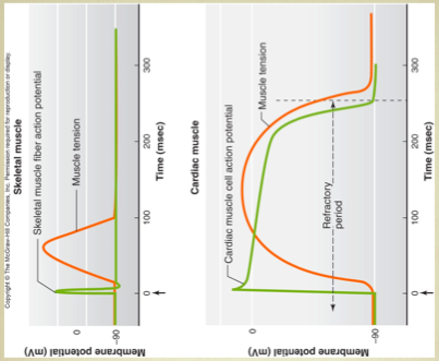
MEMBRANE POTENTIAL SKELE MUS & CARDIAC CONTRACTIONS |
|
cardiac contac are prolonged |
|
MEMBRANE POTENTIAL SKELE MUS & CARDIAC CONTRACTIONS |
|
cardiac contac are prolonged |
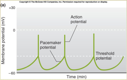
| MEMBRANE POTENTIAL (MV) |
|
- rhythmic changes in the membrane potential of smooth mus. results in rhythmic patterns of action potentials and therefore rhythmic contraction in the gut neighboring cells use gap junction to further coordinate these rhytmic contractions
|
| MOTOR UNITS |
|
-A single motor unit consists of a motor neurons and all of the mus. fibers it contols. |
| MOTOR UNITS |
|
-A single motor unit consists of a motor neurons and all of the mus. fibers it contols. |
| MOTOR UNITS ARE OF ONE TYPE |
|
| MOTORNEURON SIZE IS IMPORTANT |
|
| MUSCLE FIBER DIAGRAM |
|
| MUSCLE FIBER DIAGRAM |
|
|
| MUSCLE FIBERS HAVE DIFFERENT TWITCH KINETICS |
|
| MUSCLE TRAINING |
|
-has opposite effect of muscle distrophy - edurance training: inc. mitochondrea numbers, blood vessles around mus., and resistance to fatigue - high intensity: (str./aerobic) training stimulates inc. in fiber diameter, glycolytic enzyme levels - also inc. and more efficent neural control |
| MUSCLES ATROPHY WITH DISUSE |
|
-Destroy the motorneuron and the innervated mus. w/ atophy (shrink in diameter & exert less force) -Lack of use will cause same effect |
| MUSCLES MAY NEED LOTS OF ATP ON DEMAND |
|
| MYOSIN LIGHT CHAIN KINASE |
|
-regulates with cross-bridge cycle occurs -takes the place of troponin |
| NEUROMUSCULAR JUNCTION |
|
- 1 motor neuron may innervate many mus. fibers (cells) - this is called a motor unit - the smaller the unit = finer the control - Mus fiber membrane is in folds - motor neurons typically release acetylCholine - where the motor neurons synapses with the mus. - AP in the motor neuron cause acetylcholine release into the neuromuscular juntion - Muscle contraction follows the delivery of acetylcholine to the muscle fiber |
| OTHER DIFF. FR. SKELE MUS. |
|
- uses myo light chain kinase instead of troponin, use but don't need action potentials - Lower rate of cross-bridge cycling and longer twitch duration - myo light chain phophatase turns off cross-bridge cycle - can sustain contraction (latch state) - Innervated by autonomic nervous system |
| PATH TO PWERSTROKE |
|
-excitation-contraction coupling, -->in all 3 mus. types, Ca2+ is the trigger - signaling triggers excitatory postsynaptic potential which leads to an AC --> in mus. its called end-plate potential EEP -action potential floes into t-tubes - dihydropyride (DHP) receptor acts as a voltage sensor - Ryanodyne receptor changes conformation - ranyodynr receptor is an ion channel = opens |
| REGULATION OF CARDIAC MUS. |
|
| REGULATION OF SKELETAL MUSCLES |
|
| REGULATIONS OF THE SKELE MUS. |
|
-At the cellular level: - at the systems level: -->Increase recruitment of motor units -->small --> medium --> large motor units |
|
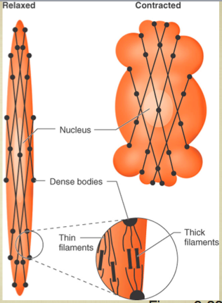
RELAXED & CONTRACTED DIAGRAM |
|
|
|
RELAXED & CONTRACTED
|
|
-Think myo based & thin actin based filaments biochem similar to those in skele. mus. fibers. --> Interact to cause smooth mus. contract. |
|
RELAXED & CONTRACTED DIAGRAM |
|
|
|
RIGORMORTIS PT. 2 |
|
-dead = no ATP made
-No ATP on myo = high affinity for actin
-stiff mus. for ~40hrs. after death |
|
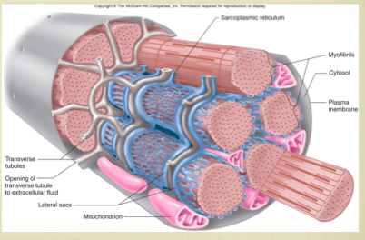
SARCOPLASMIC RETICULUM (SR) |
|
- equivalent of endoplasmic reticulum - lateral sacs contain stores of Ca2+ - transverse tubes are associated w/ lateral sacs |
| SEQUENCE OF EVENTS |
|
1. resting state = low cytoplasmic Ca2+ -->tropomyo blocks myo binding sites in actin 2. excitatory action potential triggered in mus. mem. 3. Ca2+ stores in sarcoplas. reticulum are realeased into cytoplasm 4. Ca2+ binds to troponin -> tropomyo uncovers binding sites -> myosin binds and pulls (X-bridge cycle) |
|
SEQUESNCE OF EVENTS B/T MOTOR NEURON AP AND SKELE MUS. FOBER CONTRACT. PT. 1 |
|
1. action potential is initiated and propogates to motor neuron axon terminals 2. Ca enters axon terminals through voltage-gated Ca channels. 3. Ca entry triggers release of ACh from axon terminals 4. ACh diffuses fr. axon terminals to motor end plate in muscle fiber. 5. ACh binds to nicotonic receptors in motor end plate, increasing their permiability to Na+ and K+. 6. More Na+ moves into the fiber at the motor end plate than K+ moves out, depolarizing the membrane, producing the end plate potential (EPP). |
|
SEQUESNCE OF EVENTS B/T MOTOR NEURON AP AND SKELE MUS. FOBER CONTRACT. PT. 2 |
|
7. Local currents depolarize the adjacent muscle cell plasma membrane to its threshold potential, generating an action potential that propagates over the muscle fiber surface and into the fiber along the T-tubules. 8. Action potential in T-tubules triggers release of Ca2+ from lateral sacs of sacroplasmic reticulum. 9. Ca2+ binds to troponin on the thin filaments, causing tropomyosin to move away from its blocking position, thereby uncovering cross-bridge binding sites on actin. 10. Energized myosin cross-bridges on the thick filaments bind to actin: A + M x ADP x P2 -------> A x M x ADP x P2
|
|
SEQUESNCE OF EVENTS B/T MOTOR NEURON AP AND SKELE MUS. FOBER CONTRACT. PT. 3 |
|
11. Cross-bridge binding triggers release of ATP hydrolysis products from myosin, producing an angular movement of each cross-bridge: A x M x ADP x P2 --------> A x M + ADP + P2 12. ATP binds to myosin, breaking linkage between actin and myosin and thereby allowing cross-bridges to dissociate from actin: A x M + ATP --------> A + M x ATP 13. ATP bound to myosin is split, energizing the myosin cross-bridge: M x ATP --------> M x ADP x P2 |
|
SEQUESNCE OF EVENTS B/T MOTOR NEURON AP AND SKELE MUS. FOBER CONTRACT. PT. 4 |
|
14. Cross-bridges repeat steps 10 to 13, producing movement (sliding) of thin filaments past thick filaments. Cycles of cross-bridge movement continue as laong as Ca2+ remains bound to troponin. 15. Cytosolic Ca2+ concentration decreases as Ca2+ is actively transported into sarcoplasmic reticulum by Ca2+ - ATPase. 16. Removal of Ca2+ from troponin restores blocking action of tropomyosin, the cross-bridge cycle ceases, ant the muscle fiber relaxes. |
| SHORT-TERM MUSCLE FATIGUE |
|
| SKELETAL MUSCLE FIBER TYPES |
|
| SMOOTH & SKELE MUS CONTRACT |
|
- Skele Mus.: Inc. cytosolitic Ca2+ --> Ca2+ binds to troponin on thin filaments--> Conformational change in toponin moves tropomyo out of blocking position --> Myo cross-bridge bind to actin --> cross-bridge cycle
- Smooth Mus.: Inc. cytosolitic Ca2+ --> Ca2+ binds to calmodulin in cytosol --> Ca2+ calmodulin complex binds to myo light-chain kinase --> Myo light-chain kinase uses ATP to phoyophoytate myo cross-bridges --> Phosphorylated cross-bridges bind to actin filaments --> cross-bridge cycle prod. tension & shortening |
|
SMOOTH & SKELE MUS CONTRACT PT. 2 |
|
- Ca ions play as major regulator roles in contrac. of both smooth and skele. mus., but the Ca in smooth mus. bind calmodulin activates myo light chain kinase |
| SMOOTH MUS. CAN RESPOND TO LOCAL CHANGES |
|
| SMOOTH MUS. TYPES |
|
| STEPS IN CARDIAC MUS. CONTRACT |
|
| SUMMARY OF SMOOTH MUSCLE CONTRACT. |
|
-intra. cell Ca activates calmodulin -Ca2+ : calmodulin activates myo light chain kinase - Phosporlated = myo binds actin -Unphosphorylated myo doesn't bind - myo-P can enter cross-bridge |
| THREE BASIC WAYS MUSCLES CAN MAKE ATP |
Creatine Kinase Creatine-P + ADP <------------> Creatine + ATP
|
| TITIN PROTEIN FILAMENTS HAVE SOME ELASTICITY (STRETCH) |
|
| WAYS INTRA. CELL Ca2+ LEVELS ARE REGULATED |
|
- 2 sources: 1. Sarcoplasmic Reticulum 2. extracell. Ca2+ - smaller sarcoplasmic reticulum than skele - action potential trigger release of Ca2+ fr. SR or influx fr. ions channels in plas. mem. - cell signaling can also trigger influx of Ca2+ fr. ion channels |
|
IMPORTANCE OF ATP BINDING AND HYDROLYSIS |
|
-Binding & hydrolysis is key part of process -ATP bind to myosin breaks link b/t myosin & actin -ATP-bound myosin has low affinity for actin -ATP hydrolysizes to ADP = energizes myosin & stores energy for the conformation change of powerstroke |
|
RIGOR MORTIS |
|
**shows inportance of ATP dissociat. of actin and myosin (step 3 X-bridge)**
-the gradual stiffening of skele. mus. that begins several hrs. after death -reaches a max. after ~12 hrs. -Dissapears ~40-60 hrs. after death = muscle tiss. decomposed -ATP contract. declines b/c nutrient & oxyg. supply = not avail. b/c no circulation -No ATP = no break b/t myos. & actin = thick + thin filaments still bound = thick + thin filaments can't pull past ea. other |